9.12 Anterior Thorax: Inspection
Inspection of the anterior thorax involves the following steps:
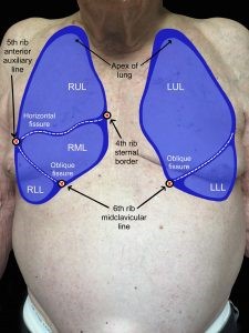
Step 1: Inspect for symmetry, chest expansion, observable deformities, masses, swelling, and shape of the thorax (see Figure 9.16a and 9.16b as a reminder for landmarks).
- Compare the left side of the thorax to the right side of the thorax. Are the clavicles and ribs on each side symmetrical upon observation? Are the ribs sloping downwards? Is the trachea and sternum midline? (note which side, if trachea is deviated [i.e., pulled to one side]). Does the chest appear elliptical in shape? (Barrel shape indicates chronic respiratory condition as commonly seen in COPD patients)
- Do you notice any deformities, masses, or swelling? (note location and describe)
- Observe the costal angle, which is the angle between the costal margins inferior to the xiphoid process. Normally, it is about 90 degrees.
- An abnormal finding is when the angle flattens out. This happens with chronic lung conditions associated with hyperinflation of the lungs (e.g., emphysema). This abnormal finding is often associated with ribs that flatten out and an anteroposterior to transverse diameter that is no longer 1:2, but rather is closer to 1:1, resembling a barrel chest.
Step 2: Inspect for skin color and skin integrity.
- Is the skin color consistent across the anterior thorax?
- Do you notice any skin discoloration or prominent vasculature?
- Do you notice any scars? If so, ask the client the cause.
Step 3: Note the findings.
- Normal findings might be documented as: “Symmetrical anterior thorax, downward sloping ribs, trachea and sternum midline, no thorax deformities, masses, or swelling, costal angle 90 degrees. Consistent skin color across anterior thorax, no discoloration”
- Abnormal findings might be documented as: “Tracheal deviation to the right side. Costal angle 170 degrees, horizontal ribs with a 1:1 anteroposterior to transverse diameter.”

Priorities of Care
Upon inspection, the findings of most concern are usually a new onset of tracheal deviation or asymmetrical lung expansion. These cues are suggestive of decreased ventilation to one side of the lungs possibly caused by pneumothorax, atelectasis, or pleural effusion. If the client is showing other signs of respiratory distress, notify the physician/nurse practitioner immediately. Otherwise, complete a primary survey (ABCCS) followed by a focused assessment of the respiratory system so that you can provide a complete report of the relevant cues to the physician/nurse practitioner.
