9.13 Anterior Thorax: Palpation
Palpation of the anterior thorax involves the following steps (see Video 9):
Step 1: Begin by using the dorsa of your hands to compare temperature bilaterally from the apex to the bases.
Step 2: While comparing bilaterally, use the finger pads of your dominant hand to palpate the anterior thorax from the apices to the base for pain, moisture, deformities, masses, swelling, or crepitus. Avoid palpating over breast tissue. You may need to ask the client to reposition their breasts so that you can palpate the thorax.
While doing so:
- Ask the client if they have any pain.
- Note any moisture or the location, size, and description of any deformities, masses, and swelling.
- Note the location and extent of any crepitus.
Video 9: Palpation of anterior thorax
Step 3: Palpate for symmetrical chest expansion by placing your hands on the patient’s chest with your thumbs meeting at the midline along the lower costal margin (around the lower ribs, just below the pectoral muscles). Your fingers should rest laterally on the chest wall, wrapping around the sides of the rib cage. Ensure your thumbs are close enough to meet at the midline, but not pressing too hard on the sternum. Make a note if the expansion is asymmetrical (one side has limited expansion, lags in expansion, or does not expand). This can happen when one lung cannot expand due to conditions that involve inflammation and air between the lungs and chest wall (i.e., pneumothorax) or partial or complete collapse of the alveoli (i.e., atelectasis).
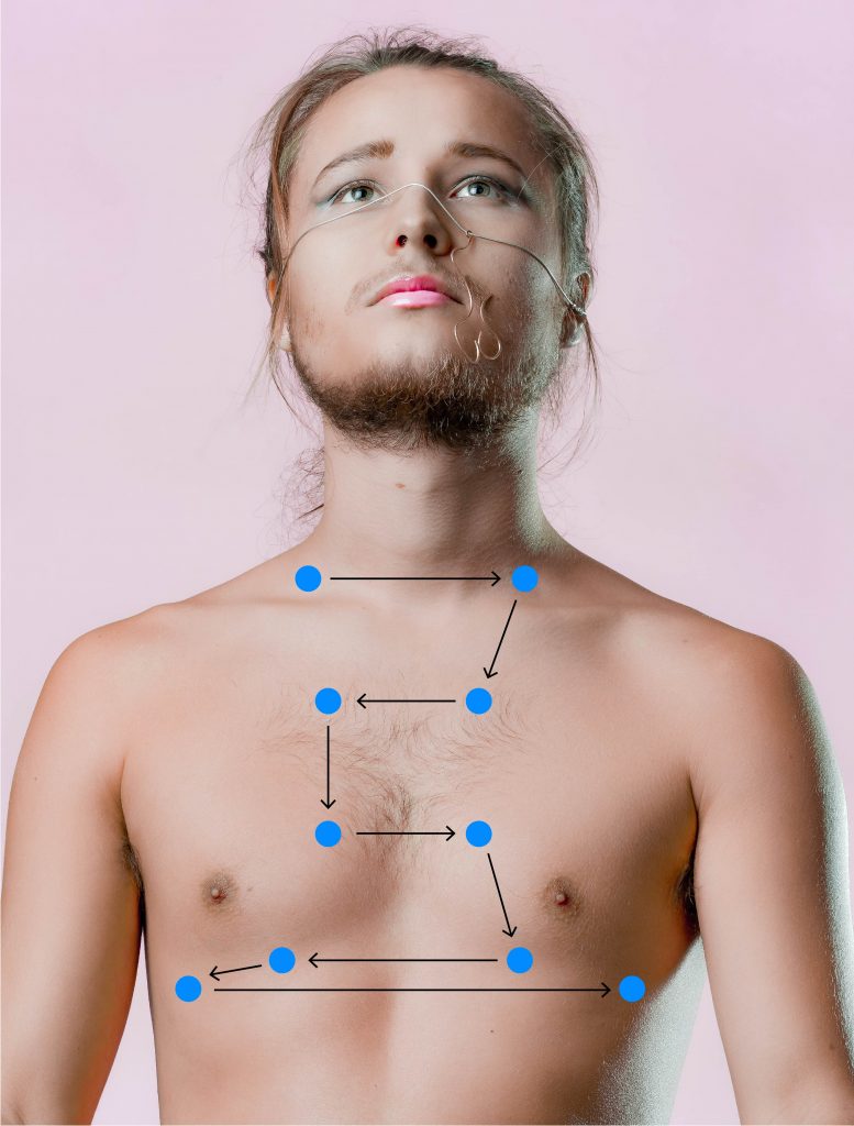
Step 4: Next, assess for tactile vocal fremitus (see Video 10).
- Place the ulnar surface of your hands or the base of the palmar aspect of your fingers on about three to five locations on the anterior thorax on each side (see Figure 9.17). Begin at the apex and move down to the base of the lungs.
- Ask the client to say “foodie” or “coin” each time you place your hands on the thorax.
- Note the equality of fremitus in comparison from the left to the right side. Remember that the vibrations will be more difficult to feel as you get closer to the bases because you are further away from the larynx.
Video 10: Palpation for tactile vocal fremitus of anterior thorax.
Step 4: Note the findings.
- Normal findings might be documented as: “Upon palpation of anterior chest wall, client reports no pain, temperature warm to touch, equal bilaterally, no moisture, swelling, masses or deformities, equal tactile fremitus.”
- Abnormal findings might be documented as: “Upon palpation of anterior chest wall, crepitus felt in neck and upper lobe area, perspiration noted.”
