9 Cell Division
Download this lab as a Microsoft Word document.
Download the lab response form (Microsoft Word document) to access the questions only.
Purpose
To understand how cells sort their chromosomes and distribute them into daughter cells during the processes of mitosis and meiosis.
Learning Objectives
At the conclusion of this exercise, students will be able to:
- Compare and contrast the processes of mitosis and meiosis.
- Identify cells in various stages of mitosis under the microscope.
- Describe the cellular processes occurring during specific stages of cell division.
- Describe chromosome structure and activity during specific stages of the cell cycle.
Why It’s Relevant
Any time an organism grows, heals, or reproduces, its cells must undergo a process of division. Understanding how each daughter cell receives a full set of genetic information so that it can function properly is crucial to understanding how life perpetuates itself. When an organism loses control of cell division, it leads to abnormalities in growth, such as cancer.
Introduction
One of the fundamental tenets of cell theory is that since the initial appearance of the first living cell on Earth, all life has come from pre-existing life. Since all living things are made up of at least one cell, this means that cells, for as long as they have existed, have been dividing to create new cells. This process is known as cell division. Cell division typically involves two important processes: the first is the proper distribution of copied genetic material (DNA) into two areas, and the second is the actual physical splitting of an existing cell into two (called cytokinesis).
Since prokaryotes, such as bacteria, have all of their DNA in a single chromosome, the process of copying DNA, organizing copies of DNA into two separate areas, and then dividing into two is relatively simple. In eukaryotes (animals, plants, fungi, and protists), however, cell division is more complicated. This is partly because eukaryotic chromosomes are contained within the nucleus of the cell, instead of floating more freely in the cytoplasm. This means that a systematic breaking down of the nucleus must occur early in cell division, and that a systematic formation of two new nuclei must occur near the end of cell division. Another reason eukaryotic cell division is more complicated is that the genetic material is typically contained on multiple, linear chromosomes, rather than just one. This requires an orderly, more structured process of organizing copied sets of DNA so that each new daughter cell produced receives a complete set of genetic instructions. Eukaryotic cell division always occurs in a highly regulated, identifiable series of stages as a result.
Cell division in eukaryotic cells always consists of the same two processes exhibited by prokaryotic cells: the organizing of the genetic material in the nucleus and subsequent cytokinesis. In eukaryotes, however, there are two different forms that nuclear organization may take. These are called mitosis and meiosis.
In mitosis, a eukaryotic cell has previously copied all of its DNA and will divide once, so that each of the two daughter cells produced receives a copy of DNA identical to its parent cell (and identical to each other). This means that mitosis always produces two daughter cells with the same amount of DNA that the parent cell exhibited. This attribute makes mitosis the process that is used anytime a eukaryotic organism is adding new cells (growing), replacing damaged cells, or engaging in asexual reproduction. Mitosis occurs in a series of recognizable stages as follows:
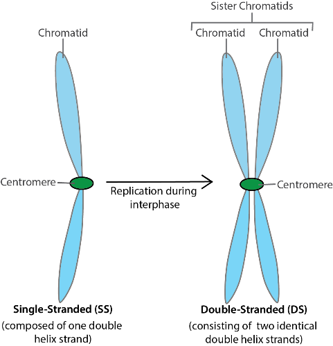
- Interphase: This stage is not technically part of mitosis but is rather its pre-phase. During interphase, cells are performing their normal functions and making preparations for cell division to occur. These include the energy-expensive process of copying all their DNA so that each single-stranded linear chromosome now exists in double-stranded form: two identical (copied) sister chromatids joined at a centromere (Figure 9.1). This is done so that once the process is complete, each produced daughter cell will contain a full set of DNA.
- Prophase: During this first stage of mitosis, many things happen in the cell at once. First, the nucleus breaks down, freeing the chromosomes (containing the DNA) into the cytoplasm. The chromosomes coil tightly onto themselves, becoming shorter and thicker (and visible with a light microscope!) Finally, a structure called the mitotic spindle begins to form. The spindle, largely formed of microtubule fibers, will attach to each of the double-stranded chromosomes and begin the process of organizing them. Over the course of prophase, two mitotic spindles are growing in length and actively moving the duplicate sets of chromosomes towards the center of the cell. Because this organization takes a little bit of time, chromosomes may appear scattered throughout the cell during this phase.
- Metaphase: This stage is achieved once all of the double-stranded chromosomes have been lined up in the center of the cell in a single row by the mitotic spindles. The spindles are now very large and originate from opposite ends of the cell. The centromere of each double-stranded chromosome is held by microtubules from each of the two mitotic spindles.
- Anaphase: Now that the double-stranded chromosomes are properly aligned, both of the mitotic spindles begin to shorten. This exerts a strong pulling force on each of the double-stranded chromosomes from opposing sides. Eventually, the chromosomes split into two at the centromere and the two sister chromatids are pulled to opposite ends of the cell (in single-stranded form).
- Telophase: The final stage of the process is telophase. Here, the sets of single-stranded chromosomes, now found at opposing ends of the cell, start to uncoil, becoming longer and thinner. At the same time, new nuclei are built around the two sets of chromosomes. Near the end of telophase, the process of cytokinesis begins somewhere in between the two sets of chromosomes. The end result is two new cells, each with a complete set of single-stranded chromosomes, genetically identical to the parent cell.
While the process of mitosis produces genetically identical daughter cells, a second version of nuclear organization of chromosomes, called meiosis, evolved to produce genetically variable offspring. While there are many similarities between mitosis and meiosis, there are three key differences:
- Meiosis always involves two cell divisions (not one, as in mitosis)
- Since there are two division events, four daughter cells are produced from a single parent cell
- Since DNA is replicated once at the start of the process and there are two division events, each of the four produced daughter cells will contain only half of the normal amount of DNA and is genetically distinct from the others
In animals, meiosis is the process used to produce egg and sperm for use in sexual reproduction. Biologists use the same stage names in meiosis as they do in mitosis, because most of the processes that occur in each stage are the same. Because cells divide twice in meiosis, the stages are designated I or II. There are some key differences, however, as outlined below.
- Interphase I: All of the same events occur as they do during the interphase that precedes mitosis. DNA is replicated so that every chromosome is in double-stranded form (consisting of two sister chromatids held together by a centromere). As in mitosis, each chromosome currently has a homologous partner, which is a “matching” chromosome of equal length and containing all of the same genes. Cells possessing chromosomes in pairs are said to be diploid (denoted by writing “2n”).
- Prophase I: All of the same events occur as they do during prophase of mitosis. The nucleus breaks down and the two mitotic spindles attach to each of the copied (double-stranded) chromosomes. In meiosis, however, pairs of double-stranded homologous chromosomes are moved towards the center of the cell, while engaging in crossing over, which means they may exchange equal length stretches of DNA, creating genetic diversity. They attach to each other, making the exchange possible. It is important to realize that homologous chromosomes always “cross” the same stretch of DNA, meaning they never lose or gain genes, but might trade versions of genes with each other.
- Metaphase I: In this stage, pairs of double-stranded homologous chromosomes are lined up in the center of the cell, in a double row. Each pair of homologous chromosomes is attached to microtubule fibers of both mitotic spindles. Since pairs of chromosomes line up independently of other pairs, there are many different arrangements possible! Along with crossing over, this independent assortment is another important source of genetic variation in egg and sperm cells.
- Anaphase I: Both mitotic spindles begin to shorten, exerting stress on each pair of chromosomes. Double-stranded homologous chromosomes break apart from their partners and are pulled to opposite ends of the cell.
- Telophase I: New nuclei are formed around each set of double-stranded chromosomes as they uncoil and cytokinesis I occurs. The end result of meiosis I is two daughter cells, each with a half set of chromosomes that are still in double-stranded form. Since homologous partners have been separated from each other in this step, the two daughter cells are no longer diploid. They are considered to be haploid (denoted by writing “n”).
- Meiosis II is essentially identical to the process of mitosis. Each daughter cell will divide again through a similar process so that its double-stranded chromosomes are split into single-stranded form. On some occasions, the start of meiosis II immediately follows the end of cytokinesis I. In others, there is a brief interlude (Interphase II), during which DNA replication does not occur as it did in the first interphase.
- Prophase II: Double-stranded chromosomes are moved towards the center of the cell in both of the two daughter cells produced by meiosis I.
- Metaphase II: Double-stranded chromosomes are lined up in a single row in the center of the cell.
- Anaphase II: Double-stranded chromosomes are split into single-stranded chromosomes at the centromere by shortening mitotic spindles on each side.
- Telophase II: New nuclei are built around the two sets of DNA in the opposing ends of both daughter cells. Cytokinesis follows, creating four daughter cells from the original cell that began meiosis I. Each has half of the number of chromosomes as the starting cell. Due to crossing over in prophase I and the random assortment of chromosomes in metaphase I, each of the four daughter cells produced is genetically unique.
Procedure
A major goal of your work in this lab is to learn to recognize the stages of mitosis described above in plant and animal cells.
To begin, obtain an Allium (onion) root tip slide and view it at scanning power (40X total magnification) under the microscope. Once you properly focus the microscope, you should see several elongated structures. These are the microscopic tips of roots of an onion plant. Since roots grow from their tips, you should be able to view many cells in the process of cell division here.
Choose one of the root tips and focus the view near its end at low (100X total magnification) and then high (400X total magnification) power. Notice that the very end of the root tip is covered by a protective root cap. These are dead cells that are constantly sloughed off as the root pushes through the abrasive soil. You will see many cells in various stages of division immediately behind (above) the root cap. On this slide, the chromosomes of the cells have been stained purple (or some other dark color) and they will appear as little sticks in each cell.
You may find that many of the cells appear to have no purple stain in them. In others, you will simply see a single dark-stained circle. These are cells that are currently in interphase. The chromosomes have not yet coiled up tightly enough for you to discern linear forms. In other cells, the linear chromosomes will be visible at one of the stages of their movement during cell division.
Look around on the slide for a good example of interphase and the four stages of mitosis and make a sketch of each of them. Keep in mind that mitosis is a process in which one stage flows into the next. You will find many examples of transitions between stages, a “late prophase” or an “early metaphase,” say. Your job is to locate a good example of each stage that cannot be confused with another.
See if you can locate a cell near the very end of telophase, starting the process of cytokinesis. You should be able to observe a new cell plate being built between the sets of daughter nuclei.
As you view the root tips, it may become obvious that some stages are better represented than others. Think of the slide you are viewing as a photograph in which time was frozen in an instant. It is logical, then, to consider that stages of mitosis that take longer than others will be well represented on the slide. The longer a stage takes, the more cells will have happened to be in that stage when the slide was made. The opposite is also true: the faster a stage occurs, the less likely it will be to “catch” a cell doing it when the slide is made.
Onion root cells have been shown to take approximately 960 minutes to complete one cell cycle (interphase–mitosis–back to interphase). Work with your lab partner to estimate the length of time that each stage of mitosis takes. Note that the onion cells are arranged roughly in a series of columns, one right next to another. One of you should focus on the root tip at 400X total magnification and look at a cell at the bottom of a column of cells. Identify the stage of mitosis that cell is in and proceed up the column, calling out the stage as you identify it. Your lab partner will make tally marks to keep track of how many instances of each stage are represented by the cells. When you reach the top cell in your view, move over one column and proceed by identifying the stages as you “read” down the next column. “Read” up the 3rd column, and so on. Continue until you have identified approximately 120 cells.
Next, switch roles with your lab partner and repeat the procedure. The second lab partner should choose a different root tip on the same slide. Pool your results together. Finally, create a mathematical way to estimate the amount of time each stage of mitosis takes based on your experimental data and the fact that the entire process takes ~960 minutes. Display your calculated results in a neat fashion. Your instructor will help you with this if needed.
Next, obtain a whitefish blastula slide. A blastula is an early stage of animal development in which the embryo consists of a hollow ball of cells. Think of an animal blastula like a root tip in plants—it is an area of very active growth where we will expect to see many cells in the process of mitosis.
Under scanning power (40X total magnification), you will see that the slide contains multiple blastulas. Choose one and view it under high power (400X total magnification). The chromosomes have been stained on the slide as they were for the onion root tip, but the chromosomes in these blastulas tend to appear smaller and are harder to see. Repeat the same procedure you did for the root tip slide—find a good example of interphase and of the four stages of mitosis and sketch each of them. Note that cytokinesis begins earlier and appears differently in animal cells than in plant cells. When you locate a blastula cell in telophase you will observe that cytokinesis by cleavage has already started to occur!
Once you have finished viewing both the onion root tip and whitefish blastula slides, you will attempt to simulate the process of meiosis at your lab bench. Head to the lab supply and obtain a set of blue-colored pipe cleaners consisting of 2 long, 2 intermediate, and 2 short pipe cleaners. These will serve to represent three different chromosomes in a cell. Obtain a matching set of red-colored pipe cleaners as well and six beads to serve as centromeres.
Interphase I
Imagine that the lab bench in front of you is the inside of a cell that is preparing to divide by meiosis. Simulate the process of DNA replication during interphase by building double stranded chromosomes out of the pipe cleaners and beads. The different pipe cleaner lengths represent different chromosomes, while the different colors of the same length represent homologous chromosomes. Begin by laying 1 long, 1 intermediate, and 1 short chromosome of each color on the bench in front of you. This hypothetical cell contains six chromosomes (human cells have 46) and is diploid, because the long, intermediate, and short chromosomes all have homologous partners of a different color. However, it is not yet ready to divide, because its chromosomes have not replicated and all six are in single-stranded form. Use pens of two different colors to make a sketch of what the chromosomes in your cell look like currently. Label your sketch “Interphase, pre-replication” and include the cell’s ploidy level (diploid or haploid).
Build six double-stranded chromosomes by combining the two pipe cleaners of the same length and color with a bead. When complete, you should have red and blue double-stranded chromosomes of 3 different lengths. Additionally, this cell is ready for division because its chromosomes have replicated themselves and are in double-stranded form. Make a second sketch of what the chromosomes look like currently. Label the sketch “Interphase, post-replication” and include its ploidy level.
Prophase I
Place each chromosome so that it is touching its homologous partner (e.g., the large red chromosome should be touching the large blue chromosome, etc.). Recall that homologous chromosomes will pair, attach to each other, and then “cross over” during Prophase I by exchanging matching portions of their chromatids during this step. We will not actually simulate crossing over in this activity, but make sure to understand that it occurs. Make a sketch of what the chromosomes look like currently and label it with the stage name and ploidy level.
Metaphase I
Move the three connected homologous partners to the exact center of your cell to simulate metaphase I. This will create a “double row” of chromosomes because the homologous partners are attached to each other. It does not matter how many of each color are on the left side or the right side of the row you form in this way. Make a sketch of what the chromosomes look like currently and label it with the stage name and ploidy level.
Anaphase I
Separate the homologous chromosomes from each other by moving those on the left of the row to the far left side of your cell, and moving those on the right in the other direction. Do not break apart double-stranded chromosomes by pulling any pipe cleaner out of its bead at this point. Make a sketch of what the chromosomes look like currently and label it with the stage name and ploidy level.
Telophase I (and Cytokinesis I)
Recall that in this stage the chromosomes uncoil and that new nuclei are formed around each set of chromosomes. The original cell now splits into two. Imagine that your lab bench now consists of two cells, each with its own set of chromosomes. Note that each of your new cells contains only 3 chromosomes, and they each no longer have homologous partners. In addition, each of the two cells is ready to divide again, because all three of its chromosomes are still in double-stranded form. Make a sketch of what the chromosomes look like currently in each of the two cells and label it with the stage name and ploidy level.
Prophase II
Each of your two cells has started the process of dividing again. The nuclei that were built in telophase I dissolve and the chromosomes move throughout the cell. Make a sketch of what the chromosomes look like currently in each of the two cells and label it with the stage name and ploidy level.
Metaphase II
Move the chromosomes in each of the two cells to the exact center of the cell. This will create a single row of three chromosomes in each cell. Make a sketch of what the chromosomes look like currently in each of the two cells and label it with the stage name and ploidy level.
Anaphase II
Break your double-stranded chromosomes by removing the pipe cleaners from each bead and moving them to opposite ends of each of your two cells. Set the remaining beads to the side of the simulation. Make a sketch of what the chromosomes look like currently in each of the two cells and label it with the stage name and ploidy level.
Telophase II (and Cytokinesis II)
New nuclei form around each of the four sets of chromosomes and the two cells split into four. Note that each cell now contains three chromosomes (1 long, 1 intermediate, and 1 short), all of which are in single-stranded form. Because crossing over occurred in prophase I, and because chromosomes assorted independently during metaphase I and II, each of the four daughter cells is genetically unique. Make a sketch of what the chromosomes look like currently in each of the four cells and label it with the stage name and ploidy level.
Alternate Procedures for Online Sections
Follow the standard methods as they are written in this lab for the onion root tip, the whitefish blastula, and simulation of meiosis (as described in the Procedure section above). Complete the same sketches on a separate document that you will submit for scoring and answer the summary questions below. Since you are not working in actual campus biology lab, however, make the following adjustments:
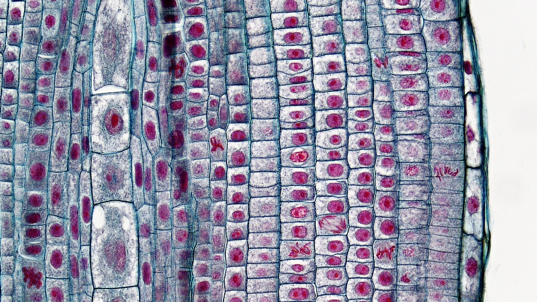
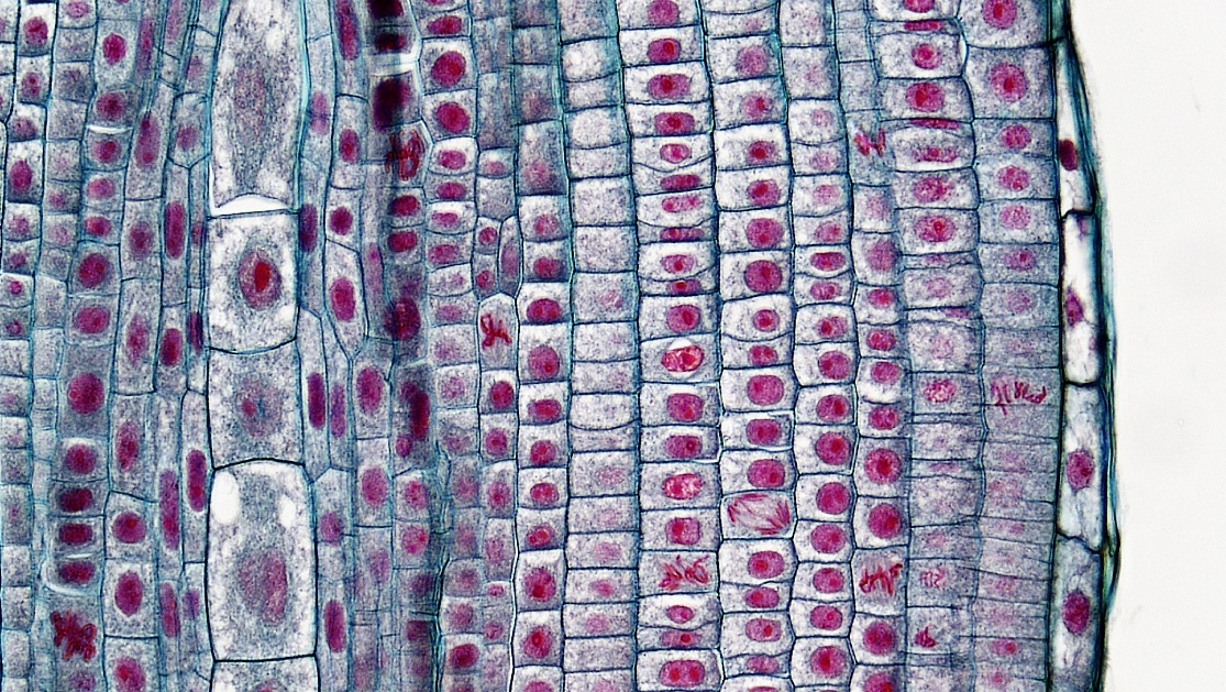
- For the onion root tip, use Figures 9.3 and 9.4 below. Locate good examples of the stages and make sketches as described in the Procedure section of this lab above. When performing the procedure for the estimate of stage length, use either Figure 9.3 or 9.4 (indicate which) and perform only 1 trial of counting approximately 120 cells.
- Skip the portion repeating the procedure for the whitefish blastula. Instead, answer the following questions comparing mitosis in plant and animal cells. Feel free to use your textbook or to conduct internet research to help you, but make sure to put answers into your own words.
-
- Describe how the process of cytokinesis differs between plant and animal cells. Use the terms “cell plate” and “cleavage” in explaining what these processes look like in these two groups of organisms.
- What is a centriole (inside a cell)? Describe the role it plays in cell division. Do animals, plants, or both use centrioles to help them divide?
- For the simulation of meiosis, you will not have access to the pipe cleaners and beads we have available in the lab. Be creative looking for replacements! You will need 12 total items to represent chromosomes, in two different colors, and of three different sizes (2 short, 2 intermediate, and 2 long in each of the two colors), and something to connect chromosomes with. The replacement could be as simple as cutting out strips of colored paper of different lengths and using tape to connect them (serving as the beads in the simulation as described in the Procedure section).
Summary Questions
- A human cell typically contains 46 chromosomes. How many chromosomes would each of the 2 daughter cells contain if the cell completed mitosis? How many chromosomes would each of the 4 daughter cells contain if the same cell completed meiosis?
- Based on your experimental results, rank the stages of mitosis (including interphase) in order from slowest to fastest. Next, suggest a plausible reason why your slowest stage takes so long.
- Use your own words to describe how the process of cytokinesis differs between plant and animal cells.
- For each of the following times during mitosis in the cell cycle, state whether the chromosomes are in single-stranded (SS) or double-stranded (DS) form:
-
- At the very beginning of interphase:
- At the end of interphase:
- At the beginning of prophase:
- At the beginning of metaphase:
- At the beginning of anaphase:
- At the beginning of telophase:

.jpg)