6 Gene Expression and Inheritance
Download this lab as a Microsoft Word document.
Download the lab response form (Microsoft Word document) to access the questions only.
Purpose
The purpose of this activity is to demonstrate gene expression and genetics through the analysis of a sickle cell anemia case study.
Learning Objectives
At the conclusion of this exercise, students will be able to:
- Identify the phenotypic differences between healthy red blood cells and those affected by sickle cell anemia.
- Explain how these phenotypic differences contribute to the symptoms of sickle cell anemia.
- Compare and contrast the phenotype and genotype of healthy and sickled red blood cells.
- Describe the molecular mechanisms underlying sickle cell anemia.
- Illustrate the genetic basis and inheritance patterns of sickle cell anemia.
- Develop a treatment plan for managing sickle cell anemia.
Why It’s Relevant
Have you ever wondered why you look or behave the way you do? Why does your smile resemble your biological mother’s, but maybe not your siblings’? Why do you share your father’s temperament? Much of this comes down to the deoxyribonucleic acid (DNA) and genes within your genome, inherited from your biological parents. All living organisms, from bacteria to animals, carry DNA, which serves as a unique identifier. This powerful macromolecule has not only deepened our understanding of species evolution but has also driven groundbreaking innovations in healthcare and agricultural practices, and has played a crucial role in advancing fairness in criminal and social justice systems.
Introduction
Deoxyribonucleic acid (DNA) is the universal language of all living organisms, providing instructions for building macromolecules, ribonucleic acid (RNA), and proteins. In turn, proteins determine the characteristics or traits of an organism. Within a specific segment of DNA is a gene, which carries the information for a particular trait. Additionally, genes can exist in different versions, known as alleles. For example, in humans, there are alleles that code for brown eyes, and a different allele that codes for blue eyes. In diploid organisms, such as humans, alleles are inherited from both biological parents—one from the mother and one from the father. When these two alleles combine during fertilization, they form the offspring’s genotype—the genetic makeup for a particular trait. This genotype is expressed as a phenotype—the physical manifestation of the gene(s) for that trait. The phenotype is brought out through the processes transcription and translation and is modified through gene regulation.
In today’s activity, you and your lab partner (if applicable) will take on the role of genetic counselors for a client with sickle cell anemia. You will apply your knowledge of gene expression and genetics to assist your patient effectively.
Part 1: Research About Sickle Cell Anemia
As a genetic counselor, your role is to research hereditary disorders and analyze development in the field of genetics, the scientific study of genes and heredity. Additionally, you are responsible for educating your clients about their condition, including its symptoms. Conduct research and answer the following questions, citing reputable sources (using the Council for Science Editors [CSE] citation format) to support your responses:
- What is sickle cell anemia?
- What are the common symptoms of the disorder?
- What protein in the red blood cell is affected by the disorder?
- What is the function of the protein?
Part 2: Phenotype of the Red Blood Cells
You obtained blood smears from both a healthy individual and your client with sickle cell anemia. Compare and contrast the phenotypes of the red blood cells.
Materials and Procedure for In-Person Courses
The lab has the following equipment for you to collect your data:
- Compound microscope
- Prepared slides of typical red blood cells and sickled red blood cells
Instructions for how to view blood smears underneath the compound microscope if you have not completed Lab 3: Microscopy
Using the compound microscope, observe and draw the two blood smears.
- Plug in the microscope and make sure the light source is on.
- Place the microscope slide labeled “Human blood smear” (blood smear from someone who does not have sickle cell) on the stage of the microscope and secure it with the slide holder.
- Rotate to the scanning objective lens (4x) to view the cells within the field of view.
- Use the coarse adjustment knob to bring the stage all the way up.
- Use the fine adjustment knob to focus and sharpen the view of the cells.
- Rotate to the 10x objective lens for low power (total magnification 100x) and use the fine adjustment knob to focus and sharpen the view of the cells.
- Rotate to the 40x objective lens for high power (total magnification 400x) and use the fine adjustment knob to focus and sharpen the view of the cells.
- Draw the image of the cells and complete the questions in Table 6.1 below.
Alternative Procedure for Online Courses
- Your instructor will provide microscope images of the blood smears.
- Draw the images of the cells and complete the questions in Table 6.1 below.
- Table 6.1. Compare and contrast the phenotypes of the blood smears.
|
|
Blood smear of person without sickle cell anemia | Blood smear of your patient |
|---|---|---|
|
Drawing of image underneath the compound microscope at high power (40x) |
|
|
|
What are the shapes of the cells? |
|
|
|
Are the red blood cells evenly distributed or clumped together in the blood smears? |
|
|

- After examining the blood smears, hypothesize how the phenotype of the sickled blood cells contributes to the symptoms of the disorder (as listed in question 2).
Part 3: Gene Expression of the Sickled Red Blood Cell Phenotype
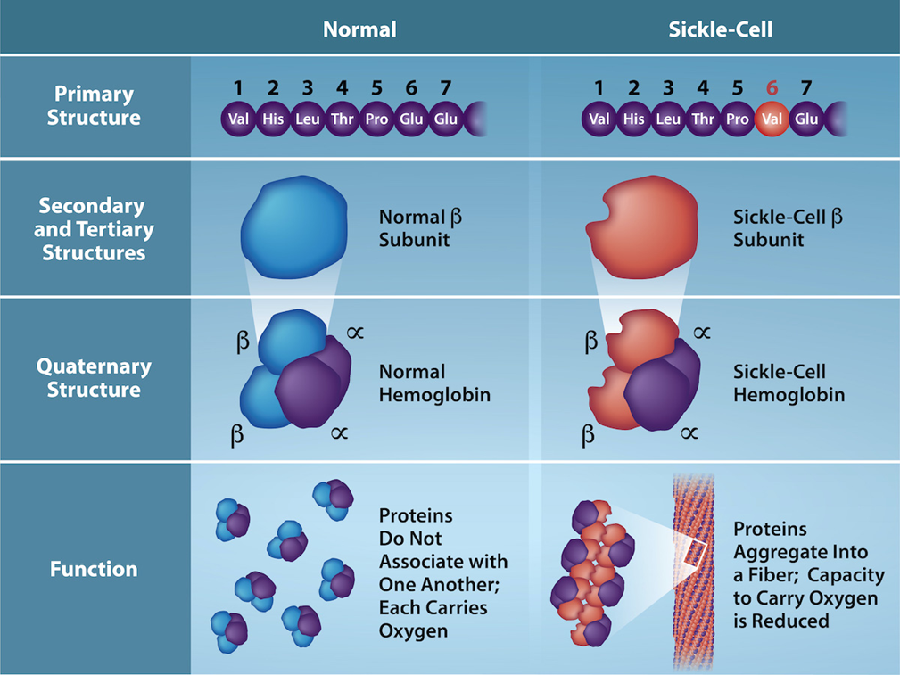
Your patient asks why their red blood cells have an unusual shape. You explain that in individuals with sickle cell anemia, the hemoglobin protein is misfolded compared to normal hemoglobin (Figure 6.1). This structure change causes the red blood cells to take on a sickled shape.
Additionally, you explain that hemoglobin is produced by a single gene in the human genome. Below is a portion of a typical hemoglobin gene sequence (Table 6.2):
|
DNA Sequence |
CAC |
CTG |
ACT |
CCT |
GAG |
GAG |
|---|
Your patient is confused, as they do not understand the connection from DNA to the protein. To help them understand, you explain the conversion from the DNA sequence into protein.
- Which molecular process does the partial hemoglobin gene (Table 6.2) go through first? What are the steps of the process?
Below you will model or draw the process of the partial hemoglobin DNA sequence in Table 6.2.
Materials and Procedure for In-Person Courses
The lab has the following equipment for you to collect your data:
- Flow of Genetic Information Kit
Instructions to model transcription using the Flow of Genetic Information Kit
- Obtain the Transcription of DNA to RNA placemat, DNA nucleotide foam pieces, and RNA nucleotide foam pieces.
- Create the typical hemoglobin DNA sequence (Table 6.2) using the DNA nucleotide foam pieces.
- String the DNA sequence through the RNA polymerase as shown in Figure 6.2.
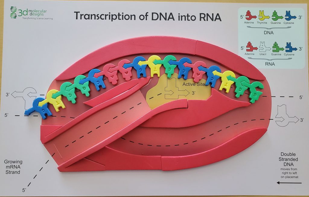
- Sprinkle free RNA nucleotides around the enzyme and complete the RNA base-pairing process on your placemat as shown in Figure 6.3.
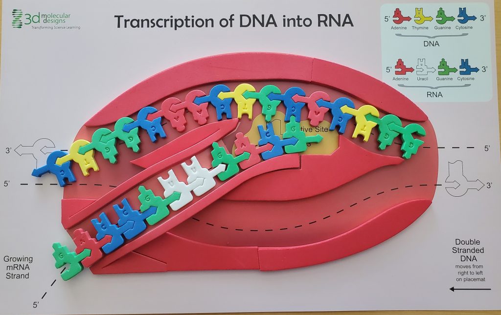
- Take a photo of your model and label the DNA template strand, the mRNA strand, and the RNA polymerase.
Alternative Procedure for Online Courses
- Draw the elongation phase of transcription for the typical hemoglobin DNA sequence (Table 6.2). Include and label the DNA template strand, the mRNA strand, and the RNA polymerase.
- Add your labeled photo or drawing of transcription of the typical hemoglobin DNA sequence below.
Once you have modeled or drawn the transcription process, write down the complementary base pairs for the mRNA sequence in Table 6.3.
- Table 6.3. Partial typical hemoglobin DNA sequence and complementary mRNA sequence.
|
DNA Sequence |
CAC |
CTG |
ACT |
CCT |
GAG |
GAG |
|---|---|---|---|---|---|---|
|
mRNA Sequence |
|
|
|
|
|
|
- Which molecular process does the partial hemoglobin mRNA sequence (Table 6.3) go through next? What are the steps of the process?
Below you will model or draw the process of the partial hemoglobin mRNA sequence in Table 6.3.
Materials and Procedure for In-Person Courses
The lab has the following equipment for you to collect your data:
- Flow of Genetic Information Kit
Instructions to model translation using the Flow of Genetic Information Kit
- Obtain the Translation of RNA to protein placement, your mRNA foam strand from the transcription part, tRNA foam pieces, and amino acid foam pieces.
- String the mRNA foam strand through the ribosomes as shown in Figure 6.4.
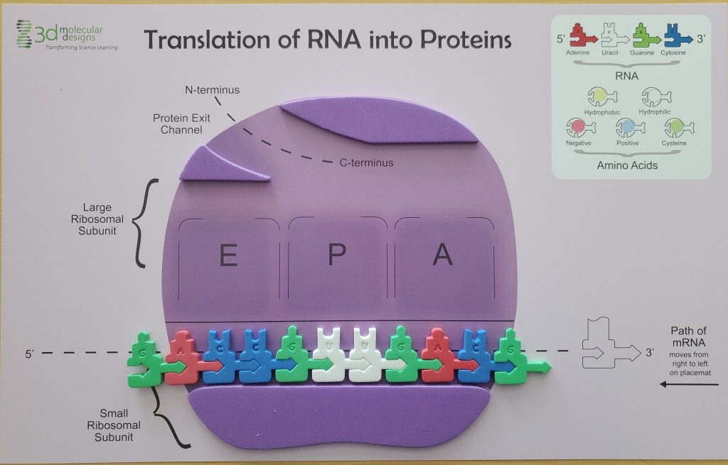
- Complete the tRNA base-pairing process on your placement as shown in Figure 6.5.

- Take a photo of your model and label the ribosome (large and small subunit), the mRNA strand, the tRNA, and amino acid sequence.
Alternative Procedure for Online Courses
- Draw the elongation phase of translation for the typical hemoglobin mRNA sequence (Table 6.3). Include and label the ribosome (large and small subunit), the mRNA strand, the tRNA, and amino acid sequence.
- Add your labeled photo or drawing of translation of the typical hemoglobin DNA sequence below.
Once you have modeled or drawn the translation process, write down the amino acid sequence complementary to the codon in Table 6.4.
- Table 6.4. Partial typical hemoglobin DNA sequence, complementary mRNA sequence, and amino acid sequence.
|
DNA Sequence |
CAC |
CTG |
ACT |
CCT |
GAG |
GAG |
|---|---|---|---|---|---|---|
|
mRNA Sequence |
|
|
|
|
|
|
|
Amino Acid |
|
|
|
|
|
|
Next, you explain to your patient that a mutation in the DNA sequence leads to the protein misfolding in sickle cell anemia, and complete Table 6.5 below.
- Table 6.5. Partial DNA sequence of sickle cell.
|
DNA Sequence |
CAC |
CTG |
ACT |
CCT |
GTG |
GAG |
|---|---|---|---|---|---|---|
|
mRNA Sequence |
|
|
|
|
|
|
|
Amino Acid |
|
|
|
|
|
|
- Compare and contrast the molecular sequences of the normal hemoglobin versus the sickled hemoglobin.
- What are the similarities?
- What are the differences? Write out the specific differences between the normal hemoglobin versus the sickled hemoglobin.
- How many amino acids were affected by the mutation, resulting in the misfolding of the hemoglobin and leading to sickle cell anemia?
Part 4: Inheritance of Sickle Cell Anemia
Now that the patient understands that sickle cell anemia is an inherited disorder, they ask why their parents don’t have the condition. You explain that sickle cell anemia is generally classified as an autosomal recessive disorder. With this in mind,
- What is the genotype of the patient? HbA is the normal allele and HbS is the mutated allele.
- If neither of the biological parents show symptoms, but your patient does, what are the possible genotypes of both parents?
- Based on the parental genotypes above, what is the probability they would have a child with sickle cell anemia? What is the genotypic ratio? What is the phenotypic ratio? Create a Punnett Square to calculate the ratios.
- If the patient decides to have child with someone that is heterozygous for the mutation, what are the odds that their children will live with sickle cell anemia?
- Construct a pedigree based on your answers to questions 16 through 19. Include the following three generations: the patient, the patient’s parents, and the patient’s grandparents.
Part 5: Treatment plan for sickle cell anemia
Now that your patient understands the biological mechanism underlying their blood disorder, they are wondering if a cure exists. Unfortunately, you must inform them that there is currently no cure for the disease. However, you can reassure them that there are promising treatments available that can help reduce the severity of symptoms. With this in mind,
- Research two different potential treatment plans, examining each at the molecular, anatomical, and physiological levels. Describe how each treatment would alleviate the patient’s symptoms and determine which of the two has a higher success rate. Be sure to cite (using Council for Science Editors [CSE] citation format) at least three reputable sources for each treatment plan to support your analysis.
References
Akinyanju, O. O. 2016. Sickle cell disease in sub-Saharan Africa: a review. National Institutes of Health. Available from: https://pmc.ncbi.nlm.nih.gov/articles/PMC4760089/
Dimitrievska, M., et al. 2024. Revolutionizing healing: Gene editing’s breakthrough against sickle cell disease. Blood Reviews. 65:17.
Genetics: A Conceptual Approach by Benjamin A. Pierce:
Pierce, B. A. 2020. Genetics: A conceptual approach. 6th ed. W.H. Freeman and Company.
Phelan J. 2021. What is life? A guide to biology with physiology. 5th ed. New York (NY): W.H. Freeman.
National Heart, Lung, and Blood Institute. 2023. Sickle cell disease. National Institutes of Health. Available from: https://www.nhlbi.nih.gov/health/sickle-cell-disease
U.S. Bureau of Labor Statistics. 2024. Genetic counselors. Available from: https://www.bls.gov/ooh/healthcare/genetic-counselors.htm#tab-2
Double-helical molecule that carries the cell's hereditary information.
Large molecule made up of small building blocks or subunits.
A single-stranded, often internally base paired, molecule that is involved in protein synthesis.
A biological macromolecule comprised of one or more amino acid chains.
A physical and functional unit of heredity, a sequence of DNA that codes for a protein.
A gene variation.
A cell, nucleus, or organism containing two sets of chromosomes (2n).
The process where a sperm units with an egg (ovum) which results in a zygote.
The genetic makeup for a particular trait in an organism.
The observable trait expressed in an organism.
The process through which messenger ribonucleic acid (RNA) forms on a template of deoxyribonucleic acid (DNA)
The process through which ribonucleic acid (RNA) directs the protein's formation.
The process that induces or represses the expression of a gene
A genetic disorder that affects the shape of red blood cells, and their ability to transport oxygen and move through capillaries.
The process that produces function protein or ribonucleic acid (RNA) from a gene.
The study of genes, genetic variation, and heredity in organisms.
The passing of traits from one generation to another.
A small (7–8 μm) biconcave cell without mitochondria (and in mammals without nuclei) that is packed with hemoglobin, giving the cell its red color; transports oxygen through the body
A monomer of nucleic acids; contains a pentose sugar, one or more phosphate groups, and a nitrogenous base
An enzyme that produces RNA that is complementary to the DNA template
A catalyst in a biochemical reaction that is usually a complex or conjugated protein.
A RNA that carries activated amino acids to the site of protein synthesis on the ribosome.
A monomer of a protein.
A cellular structure that carries out protein synthesis.
Three consecutive nucleotides in mRNA that specify the insertion of an amino acid or the release of a polypeptide chain during translation
A variation in the nucleotide sequence of a genome.
A description of a genetic trait or condition that can be passed down from parent to child. The trait is recessive located on a autosomal chromosome.
A visual representation of a cross between two individuals in which the gametes of each individual are denoted along the top and side of a grid, respectively, and the possible zygotic genotypes are recombined at each box in the grid
Having two different alleles for a given gene on the homologous chromosome.
